Hyperacute T Waves vs. Peaked T Waves vs. Normal T Waves
Key takeaways
- Normal T waves are usually smooth and asymmetric, and they may be prominent without being pathologic.
- Hyperkalemic peaked T waves are typically tall, narrow, pointed, symmetric, and diffuse.
- Hyperacute T waves are usually broad-based, bulky, symmetric, and regional, often representing early acute coronary occlusion.
- The implications differ substantially: Normal T waves may need no intervention, peaked T waves may signal a potassium emergency, and hyperacute T waves may signal an occlusion emergency.
- Morphology, lead distribution, serial change, and patient context are more informative than T-wave height alone.
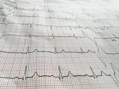
Tall T waves can be easy to notice and surprisingly hard to interpret. On an electrocardiogram (ECG), a prominent T wave may represent a normal repolarization variant, early hyperkalemia, or acute coronary occlusion. The challenge is that all three can look “big” at first glance, but they do not carry the same urgency. For clinicians, the key is not simply deciding whether the T wave is tall. The key is recognizing the morphology, distribution, associated ECG findings, and clinical setting that make the tracing benign, worrisome, or immediately actionable.
Why this distinction matters
Confusing hyperacute T waves with hyperkalemic peaked T waves can delay reperfusion in a patient with acute coronary occlusion. Confusing a benign prominent T wave with either of those abnormalities can lead to unnecessary alarm. At the same time, dismissing true peaked T waves as a normal variant can be dangerous because hyperkalemia may progress rapidly to conduction delay, bradyarrhythmia, ventricular arrhythmia, and cardiac arrest. ECG interpretation in this setting is high stakes because the tracing often provides the first clue that something time-sensitive is happening.
What a normal T wave usually looks like
A normal T wave generally has a smooth, asymmetric contour. In most leads, the upslope is more gradual, and the downslope is steeper. The wave is not sharply pointed, and it usually appears proportionate to the preceding QRS complex. Prominent T waves can still be normal, especially in younger adults and in some precordial leads, but a normal prominent T wave does not usually look narrow, needle-like, or abruptly tented. It also should not be accompanied by new PR prolongation, QRS widening, or evolving ST-segment change. The most reassuring clues are stability over time and a clinical context that does not suggest electrolyte disturbance or acute ischemia.
What peaked T waves look like
Peaked T waves — in the classic hyperkalemic sense — are tall, narrow, pointed, and fairly symmetric. Many clinicians describe them as tented. They often appear diffusely rather than in one clear anatomic territory. In early hyperkalemia, the T waves may be the first ECG abnormality to appear. As potassium rises further, additional findings can develop, including PR prolongation, flattening or loss of the P wave, QRS widening, and eventually a sine-wave pattern in severe cases. The T wave itself often looks slimmer and more sharply pointed than the bulky T wave seen in acute ischemia.
What hyperacute T waves look like
Hyperacute T waves are the early ischemic T waves associated with acute coronary occlusion. They are typically broad-based, large, and symmetric, but not sharply tented. Compared with hyperkalemic T waves, they tend to look fuller and heavier, with a wider base. They are also more likely to be localized to leads representing a vascular territory rather than appearing diffusely across the ECG. Hyperacute T waves may precede obvious ST-segment elevation, and they often evolve on serial tracings. That dynamic behavior is one of the most useful clues that the process is ischemic rather than metabolic.
Hyperacute T waves vs. peaked T waves
This is the distinction that matters most at the bedside. Hyperkalemic peaked T waves are classically narrow-based and diffuse. Hyperacute ischemic T waves are broad-based and regional. Hyperkalemia often brings associated conduction abnormalities as severity increases, including PR prolongation and QRS widening. Acute coronary occlusion is more likely to produce evolving ST-segment change, reciprocal change, ischemic symptoms, and a distribution that fits coronary anatomy. Both patterns can look tall and symmetric, but they do not have the same geometry. One looks tented. The other looks swollen and broad.
Normal T waves vs. peaked T waves
The difference between a normal prominent T wave and a pathologic peaked T wave is often more about shape and context than absolute height. A normal T wave may be tall but still smooth, slightly asymmetric, and proportionate to the QRS complex. A hyperkalemic peaked T wave is more likely to be sharply pointed, symmetric, and present in multiple leads. It also becomes much more concerning when paired with kidney disease, missed dialysis, potassium-retaining medications, metabolic acidosis, weakness, or bradycardia. A tracing that appears unchanged from prior ECGs is more reassuring than one that is clearly new.
What each type of wave implies clinically
Normal T waves
Normal T waves imply physiologic ventricular repolarization. Even when they are somewhat prominent, they are not necessarily pathologic if the patient is asymptomatic, the morphology is not sharply abnormal, and the ECG is stable over time. The implication is usually that no emergency intervention is needed solely on the basis of T-wave appearance. Clinical correlation still matters, especially in patients with chest pain or renal disease, but the tracing itself does not force an urgent differential when the pattern is clearly baseline and benign.
Peaked T waves
Peaked T waves imply possible hyperkalemia until proven otherwise, especially when they are diffuse and narrow-based. Their presence should prompt rapid consideration of serum potassium measurement, medication review, renal function assessment, and immediate treatment planning when the clinical picture supports true hyperkalemia. The implication is urgency because hyperkalemia can deteriorate into life-threatening conduction disturbance and malignant arrhythmia. At the same time, the ECG is imperfect — serious hyperkalemia can exist without classic peaked T waves, and the degree of ECG change does not always track cleanly with the serum potassium level.
Hyperacute T waves
Hyperacute T waves imply possible acute coronary occlusion, often at an early stage before the tracing meets classic STEMI criteria. Their main implication is that clinicians should not wait for dramatic ST elevation if the overall ECG and clinical picture point toward occlusive myocardial infarction. These T waves are important because they can be an early reperfusion-era warning sign that myocardium is at risk now, not later. Serial ECGs, symptom timing, reciprocal changes, and urgent ischemic evaluation become central when this pattern is suspected.
Detailed morphology: What to look for lead by lead
When deciding whether a T wave is normal, peaked, or hyperacute, it helps to ignore the label at first and describe what is actually on the page. Is the wave narrow or broad? Is it diffuse or regional? Is it proportionate to the QRS, or does it look oversized relative to the preceding complex? Are the ST segments changing? Is the tracing evolving compared with a prior or a repeat ECG? Hyperkalemia tends to create a diffuse repolarization abnormality with progressive conduction effects. Acute ischemia tends to create a territorial abnormality with dynamic change. Normal variants tend to remain stable, proportionate, and unaccompanied by new interval abnormalities.
Common interpretation traps
One common trap is using the word “peaked” for every tall T wave. In clinical ECG language, that shorthand can blur the important distinction between a narrow hyperkalemic T wave and a broad hyperacute ischemic T wave. Another trap is assuming that hyperkalemia must always produce dramatic tented T waves. In reality, ECG findings correlate imperfectly with potassium level, and some patients with dangerous hyperkalemia have only subtle changes or no classic changes at all. A third trap is ignoring distribution. Diffuse abnormalities suggest a systemic process such as hyperkalemia. Regional abnormalities suggest ischemia until proven otherwise.
A practical bedside framework
A useful approach is to ask four questions. First, is the T wave narrow and tented, or broad and bulky? Second, is the pattern diffuse or localized to a vascular territory? Third, are there associated findings such as PR prolongation, QRS widening, reciprocal change, or evolving ST-segment abnormality? Fourth, does the patient’s story fit hyperkalemia, acute coronary syndrome, or a benign baseline pattern? Framing the ECG this way helps separate waveform recognition from diagnosis and reduces the risk of anchoring too quickly on T-wave height alone.
Frequently Asked Questions
Are hyperacute T waves and peaked T waves the same thing?
No. Both can look tall and symmetric, but hyperacute T waves are usually broad-based and regional, while hyperkalemic peaked T waves are classically narrow-based and diffuse.
Can a normal ECG have tall T waves?
Yes. Some patients, especially younger adults, can have relatively prominent T waves as a normal variant. Stability over time and the absence of concerning associated findings help support that interpretation.
Do peaked T waves always mean hyperkalemia?
No, but diffuse narrow tented T waves should strongly raise that concern. The differential still depends on morphology, distribution, and clinical context.
Do hyperacute T waves always come with ST elevation?
No. They may appear before obvious ST elevation and can be an early sign of acute coronary occlusion.