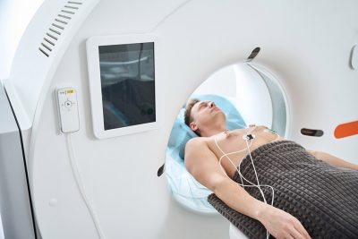
Routine CCTA Imaging of Left Main PCI Patients Falls Short in Randomized Trial
October 1, 2025
By Jeffrey Zimmet, MD, PhD
Synopsis: In this randomized trial of patients undergoing left main percutaneous coronary intervention, routine surveillance coronary computed tomography angiography six months after intervention did not reduce the composite endpoint of all-cause death, myocardial infarction (MI), unstable angina, or stent thrombosis at 18 months, but was associated with fewer spontaneous MIs and more imaging-triggered revascularization procedures.
Source: D’Ascenzo F, Cerrato E, De Filippo O, et al. Computed tomography angiography or standard care after left main PCI? J Am Coll Cardiol. 2025; Aug 6. doi: 10.1016/j.jacc.2025.07.060. [Online ahead of print].
What imaging surveillance do patients need following left main percutaneous coronary intervention (PCI)? In the past, routine invasive coronary angiography two to six months after intervention was considered standard of care after left main stenting, but this recommendation was removed with a guideline update in 2009, and the 2011 American College of Cardiology (ACC)/American Heart Association (AHA)/Society for Cardiovascular Angiography & Interventions (SCAI) guidelines formally omitted routine follow-up angiography after PCI for left main disease as the result of evidence from clinical trials (such as SYNTAX) showing good intermediate-term outcomes.
Historically, left main PCI has been associated with higher risks of adverse cardiovascular outcomes relative to other coronary segments, despite the emergence of newer-generation drug-eluting stents. Observational studies had linked routine angiographic follow-up with long-term clinical benefit, but randomized trials in broader PCI populations showed no reduction in myocardial infarction or mortality when routine invasive or ischemia-guided surveillance was employed. Critically, these prior trials contained only a small subset of patients with left main or complex bifurcation disease.
In recent years, coronary computed tomography angiography (CCTA) has emerged as an accurate and minimally invasive tool for detecting stent patency and restenosis, including in left main segments. Its diagnostic yield in these larger proximal vessels rivals invasive angiography, offering benefits in safety and cost. Despite these advantages, the effect of routine CCTA-guided surveillance on hard clinical outcomes in left main patients post-PCI had not been directly examined until the present PULSE trial — an international, multicenter, randomized controlled study.
The PULSE trial enrolled 606 consecutive patients who underwent PCI for unprotected left main coronary artery disease using second-generation drug-eluting stents, randomized 1:1 either to routine CCTA follow-up at six months or standard symptoms/ischemia-driven care. Follow-up continued for 18 months, with a composite primary endpoint comprising all-cause death, spontaneous myocardial infarction, unstable angina, or definite/probable stent thrombosis. Secondary outcomes included target-lesion revascularization (TLR) and individual composite endpoint components.
Baseline demographic and procedural characteristics were well-balanced between the two groups; notable risk factors, such as diabetes mellitus (roughly 25% of patients), hypertension, hypercholesterolemia, and previous cardiac events, were common. A majority of patients had complex left main stenosis involving the distal bifurcation and underwent provisional stenting, with about 70% receiving intracoronary imaging (mainly intravascular ultrasound), reflecting contemporary best practices.
Routine CCTA was completed in nearly 90% of those assigned, with scans rated as diagnostic per established guidelines. The CCTA and control groups had similar rates of the composite primary outcome (11.9% vs. 12.5%; hazard ratio [HR], 0.97; 95% confidence interval [CI], 0.76-1.23; P = 0.80), indicating no overall reduction in major adverse cardiovascular events (MACE). However, the rate of spontaneous myocardial infarction was lower in the CCTA arm (0.9% vs. 4.9%; HR, 0.26; 95% CI, 0.07-0.91; P = 0.004), a statistically significant reduction with a number needed to treat of 25 for the prevention of one myocardial infarction in this high-risk population.
Secondary analyses showed that rates of overall target lesion revascularization (TLR) (10.2% vs. 7.6%), clinically driven TLR (5.3% vs. 7.2%), and non-TLR events did not differ significantly. Notably, imaging-triggered revascularization was far more common in the CCTA arm (4.9% vs. 0.3%, HR, 7.7; P = 0.001), reflecting increased detection and intervention for silent stenosis and in-stent restenosis (ISR). However, this did not translate to superior composite outcomes. Subgroup analyses found no statistically significant effect modification by common clinical or anatomical variables. Landmark and as-treated analyses confirmed the persistence of the reduction in spontaneous myocardial infarction with CCTA surveillance.
The authors concluded that, among patients treated with second-generation drug-eluting stents for unprotected left main coronary disease, routine CCTA at six months does not reduce the composite rate of death, spontaneous myocardial infarction, unstable angina, or stent thrombosis over 18 months of follow-up compared to standard symptoms/ischemia-guided care. Nonetheless, they reported that routine CCTA surveillance is associated with a significant reduction in spontaneous myocardial infarction, primarily through early detection and preemptive intervention for ISR or de novo stenoses that might otherwise remain subclinical. The authors suggested that CCTA-guided surveillance may be beneficial in patients with complex left main anatomy or other high-risk features, but additional research in these subgroups is warranted.
Commentary
For practicing cardiologists, this study addresses a longstanding question in the management of patients after left main PCI: Should routine imaging, specifically CCTA, be used to monitor for ISR and guide subsequent care? In day-to-day practice, most post-PCI surveillance is driven by symptoms or noninvasive ischemia testing, not routine anatomical imaging.
The most important takeaway is that routine CCTA surveillance after left main stenting with second-generation drug-eluting stents does not confer a reduction in the composite MACE endpoint, which aligns with previous data showing limited utility for routine invasive or ischemia-driven follow-up in stable PCI populations. Although the reported reduction in spontaneous myocardial infarction with CCTA-guided follow-up appears clinically meaningful, this result should be considered hypothesis-generating only. The trial’s relatively low event rate necessitates cautious interpretation and further study.
These results suggest that routine anatomical surveillance using CCTA can increase earlier detection of silent ISR and new stenotic lesions. Importantly, this comes at the expense of increased imaging-triggered interventions, which may not improve overall survival or reduce combined cardiovascular events. Consideration of routine imaging in specific high-risk subsets remains a tempting but unproven strategy. Note that current U.S. guidelines recommend against routine testing with CCTA or stress testing in the absence of a change in clinical or functional status.
Ultimately, the PULSE trial strengthens the evidence against universal routine imaging after left main PCI but highlights the potential of CCTA to reduce spontaneous myocardial infarction in high-risk patients. U.S. cardiologists should interpret these findings in light of patient-specific risk, resource use, and evolving guidelines, refraining from blanket adoption of routine imaging while recognizing its possible utility in targeted scenarios.
Jeffrey Zimmet, MD, PhD, is Associate Professor of Medicine, University of California, San Francisco; Director, Cardiac Catheterization Laboratory, San Francisco VA Medical Center.