
Common Foot Problems in Primary Care
January 1, 2026
AUTHORS
Kevin F. Sunshein, DPM, Fellow, American College of Foot and Ankle Surgeons; CEO, Sunshein Podiatry Associates, Centerville, OH
Carl Braunschweiger, DPM, American Board of Foot & Ankle Surgery; Diplomate, American Board of Podiatric Medicine; Sunshein Podiatry Associates, Centerville, OH
Executive Summary
Foot problems are a common complaint to primary care physicians and often mirror the patient’s general health, such as autoimmune disorders, endocrine disorders, neuropathies, and vasculopathies.
- Skin conditions include hyperkeratoses, verrucas, fungal infections, onychomycosis, and onychocryptosis.
- Painful and/or deforming conditions include heel pain, Baxter’s neuritis, retrocalcaneal heel pain, hallux valgus, Achilles tendinitis, plantar fasciitis, hammertoes, Morton’s neuroma, and metatarsalgia.
- Because the foot is the foundation for the rest of the body, proper footwear, patient education, and judicious surgical intervention can alleviate a lifetime of problems.
Introduction
Foot problems are a common chief complaint to primary care physicians and can present in many ways. They can be the first sign of more serious medical problems. The feet mirror one’s general health. Autoimmune disorders, endocrine disorders, neuropathies, and vasculopathies can present initially as symptoms in the lower extremity.
Anatomy of the Foot
The foot contains 26 bones, 33 joints, 107 ligaments, and 19 muscles.1 A basic review of foot anatomy will provide physicians with a good perspective on the foot’s complexity. The foot is divided into three different regions: forefoot, midfoot, and rearfoot. Tendons control the orientation of various joints to work together and propel the body forward.2
The hindfoot (or rearfoot) is most proximal and consists of the talus and calcaneus. The articulation point between talus and calcaneus is referred to as the subtalar joint, which consists of three facets that allow for rearfoot inversion and eversion. Tendon attachment sites, such as the Achilles tendon on the posterior surface of the calcaneus, are common sites of inflammation when excessive or abnormal motion is present. This can present as a pathology such as Achilles tendinitis, posterior tibial tendinitis, or tendinitis of any other tendon, and is not always isolated to one complaint. Likewise, the plantar aponeurosis is extremely susceptible to injury, usually at the medial attachment to the plantar calcaneal tuberosity.2
The midfoot consists of the navicular, cuboid, and three cuneiforms. The articulation between the rearfoot and midfoot is referred to as the Chopart joint, which includes the talonavicular and calcaneocuboid joints. These bones contribute to the stability of the foot through two planes that work together during various phases of gait to absorb shock and transfer load throughout the body.
The forefoot is the most distal aspect of the foot. It includes the five metatarsals and their respective digits. The forefoot further divides into the medial and lateral columns. One may refer to each metatarsal and its respective phalangeal bones as a “ray.” The medial first three rays together are referred to collectively as the medial column. The remaining lateral two rays are referred to collectively as the lateral column. The hallux has two phalanges and only one interphalangeal joint, whereas the lesser four toes have three phalanges and two interphalangeal joints.
Muscles and tendons are predominately responsible for coordinated movement of the pedal osseous structures and provide dynamic stabilization throughout standing and walking/running activities.3
Common Skin Conditions of the Foot
Hyperkeratoses, or calluses, are abnormal thickening and hardening of the skin from excessive pressure or friction on an area of the epidermis, specifically the stratum corneum. These lesions appear to be diffuse or can be nucleated with a central core of skin embedded in the center of the lesion. This skin is avascular but may be infiltrated with nerve fibers. If the lesions are on the toes either dorsally or interdigitally, they usually are referred to as hard “corns” or heloma durum. A hard corn usually is on the dorsal aspect of the interphalangeal joints or the sides of the joints and results from pressure of a prominent joint on a shoe. Soft corns, or heloma molle, frequently are between the fourth and fifth toes in the sulcus and result from pressure between two prominent joints or a prominent bone rubbing on adjacent skin.4
Treatment of these lesions is accomplished via sharp debridement with surgical blades and topical keratolytics. It is important to understand the underlying etiology of callus and corn formation for long-term symptom relief. Appropriate counseling on shoe type usually is necessary. Accommodative padding of the digits can prevent frequent recurrence of these lesions. If conservative treatment fails, surgery is required to address the underlying bony pathology. Excessive pressure on underlying skin can produce ulcerations from calluses and does not always happen in a neuropathic setting. A simple flexor tenotomy can be performed to release the pressure on the toe by releasing the contracted flexor tendon and rebalancing both the intrinsic and extrinsic tendons and muscles on the toe.5 Flexor tenotomy is not effective for rigidly contracted digits, but it effectively prevents re-ulceration in flexibly contracted digits. This can be performed easily in the office setting.
Verruca
Warts on the foot are caused by the human papillomavirus (HPV). They are the most common viral infection of the skin, affecting 7% to 10% of the general population. Different viral strains produce varying wart presentations in individual areas. Verruca plantaris refers to warts located on the plantar surface of the foot. Verruca vulgaris refers to lesions on the other surfaces of the foot, including the toes. This common skin infection affects children, adolescents, and young adults, and can occur in older adults as well. Prevalence is equal in both sexes.6 Risk factors include immunosuppression, repeated trauma to the skin, swimming in public pools, use of communal showers, contact with HPV-infected individuals, and excessive sweating.6
Signs and symptoms include single or multiple papules on the skin varying in size from 2 mm to 20 mm. The lesions may occur as a cluster called mosaic warts. The lesions can be painful or painless to direct pressure but often are painful on lateral compression. They may appear to be white dots or have dark spots indicating capillary infiltration. Inspection of the skin under magnification (dermoscopy) often is beneficial in determining the difference between a wart and a callus. Diagnostic features include absent skin lines, capillary bud formation, and pain with lateral compression. Differentials include molluscum contagiosum, seborrheic keratosis, and squamous cell carcinoma. If left untreated, verrucae can progress to verrucous carcinoma. Skin biopsy is the gold standard for diagnosis and is recommended for chronic warts.7
Treatment modalities are numerous and can be time-consuming, costly, and painful. Treatments include surgical excision, cryotherapy, laser ablation, immunomodulatory agents, topical keratolytics (salicylic acid), and intralesional injections (bleomycin).8 Some evidence suggests that oral cimetidine exhibits immunomodulatory activity through increased mitogen-induced lymphocyte proliferation and inhibition of T-cell function at histamine receptor sites.9 No one specific treatment works best, and many warts may be recalcitrant to any therapy. Treatment can last weeks to many months, and there is a risk of recurrence and spread.
Swift (Microwave) Treatment of Warts
In many cases, warts can persist despite multiple modalities of treatment. Recalcitrant warts often can be associated with the development of more malignant skin pathologies, such as squamous cell carcinomas. Recently, focused microwave technology has provided promising results.10 Microwaves are a form of non-ionizing radiation between 300MHz and 300 GHz. The application of focused microwave energy increases skin temperature to a hyperthermic range. This promotes apoptosis of keratinocytes. This novel technology also has been shown to promote adaptive immunity against HPV-infected tissue. Several advantages to microwave therapy are noted, and include more targeted treatment of tissue, short-lived pain with no scarring, and no requirement of dressing application. Additionally, unlike laser ablation, there is the absence of aerosolized viral particles.
Fungal Skin Infections
Tinea pedis is a common fungal infection of the foot. It is manifested as a pruritic, erythematous, scaly eruption of the foot. Depending on its location, it has three variant forms: moccasin or dry tinea pedis, interdigital, and vesiculobullous type. The most common tinea pedis infections are caused by dermatophytic fungi including Trichophyton spp., Epidermophyton spp., and Microsporum spp. of the dry tinea pedis presentation.11
Tinea pedis commonly occurs secondary to the moist environment present within occlusive shoe gear. It can be misconstrued as atopic dermatitis or allergic eczema. Tinea pedis commonly responds to the application of topical antifungal medications such as ciclopirox, terbinafine, econazole, or ketoconazole creams; however, it does demonstrate a high rate of recurrence. Therefore, a comprehensive treatment plan, including addressing environmental factors, is paramount in preventing self-inoculation.
Onychomycosis
Tinea unguium (onychomycosis) is a fungal infection on the matrix, plate, or nail bed, commonly associated with tinea pedis. The typical presentation of fungal-infected nails include abnormal thickening with lysis; yellow, white, or brown discoloration; malodor; and hyperkeratosis of the underlying nail bed. The most common cause is a dermatophyte. Non-dermatophyte molds and Candida species are less common. Onychomycosis is more prevalent in older populations, and men are affected more commonly than women.12 Toenails are involved more frequently than fingernails.12
The most common treatment for tinea pedis is a topical medication. The two main groups of antifungal therapy are azole and allylamine medications. Azoles are fungistatic and include ketoconazole and clotrimazole. Allylamines are fungicidal and include naftifine and terbinafine. The advantage of using an allylamine is the shorter duration of treatment.
The most effective treatment of onychomycosis is an oral antifungal agent, such as terbinafine or itraconazole.13 The duration of treatment usually is 90 days, either with daily intake or through pulse dosing. Concomitant use of topical antifungal medication for long duration may prevent recurrences, although there is a high recurrence rate.
Onychocryptosis (Ingrown Toenails)
Ingrown toenails are the most common nail problem.14 Ingrown toenails occur from improper trimming, tight or ill-fitting shoes, blunt trauma, repetitive trauma (especially that sustained by athletes), and hereditary nail shapes, such as pincer nails. They occur most commonly in those 15 to 40 years of age, but patients of all ages can experience this condition. Studies are conflicting regarding the incidence in males vs. females.14
Ingrown toenails can be infected or non-infected and occur most frequently in the great toes. The usual presentation is progressive pain along the medial or lateral nail border with periungual inflammation and edema. In later stages, purulent discharge and formation of granulation tissue is noted.
In the early presentation, cutting the distal offending nail border may relieve pain, but more progressive conditions require surgical excision of the offending nail border under a local anesthetic. If the nail is not infected, a permanent partial nail resection (partial matricectomy) can be performed to prevent recurrence of the ingrown toenail. Typically, destruction of the nail root is performed using 85% phenol or 10% sodium hydroxide. Other surgical methods are available, but chemical matricectomy is one of the easiest and most economical procedures.15
In the presence of severe periungual inflammation and drainage, oral antibiotics can alleviate clinical signs of infection but seldom cure this problem, since an ingrown toenail is a foreign body reaction. Staphylococcus aureus is the most common pathogen causing paronychial infections.14,15
A partial or total nail avulsion is recommended in this situation. This procedure does not destroy the nail root. Under local anesthesia, the offending border is split back under the hyponychium with a nail splitter, and the nail spicule is removed from the soft tissue borders. Any granulation tissue present is sharply excised. A compression dressing is applied and changed daily until the infection and inflammation have resolved, usually within 48 to 72 hours. Soaking the foot in Epsom salt twice daily for 10 to 15 minutes will help resolve the inflammation. Postsurgical oral antibiotics are indicated, depending on the severity of the inflammation of the affected area.
Heel Pain
Up to 15% of all foot complaints are related to heel pain.16 Plantar fasciitis is defined as inflammation of the plantar fascial tissue at the plantar aspect of the heel and is characterized by intense pain localized to the inferior portion of the calcaneus, most commonly along the plantar medial tuberosity. Any portion of the plantar fascia can be affected. The plantar fascia is a thick, broad, and dense band of longitudinally arranged collagen fibers that begins as an attachment to the anterior aspect of the calcaneal tuberosity.17 The plantar fascia is divided into three bands: medial, central, and lateral. Heel spur formation has been associated with the proximal plantar fascia, but the spur actually is deep to the plantar fascia and lies within the origin of the flexor digitorum brevis muscle. Most often, heel spurs exist without producing symptoms, although in some patients, heel spurs may become a source of symptoms.18 (See Figures 1 and 2.)
Figure 1. Image Showing Plantar and Posterior Spur
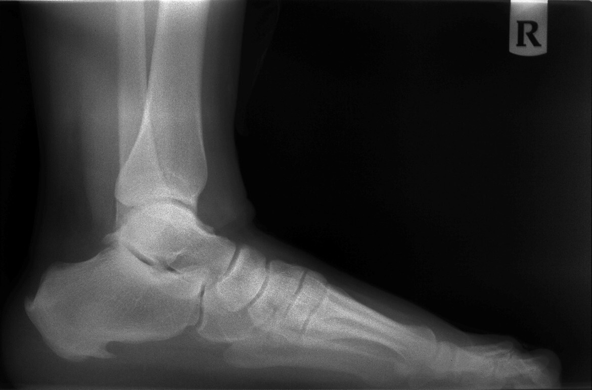
Figure 2. Image Showing Posterior Spur
Conventional plantar fasciitis is consistent in its initial presentation. Signs and symptoms include intense pain along the bottom of the heel with initial weight-bearing in the morning, commonly referred to as post-static dyskinesia.17 The pain typically subsides after a few steps, until the plantar fascia and plantar intrinsic muscles stretch out, but the pain frequently progresses throughout the day with prolonged weight-bearing. In severe cases, associated signs and symptoms include heel edema and thickening of the plantar heel pad. Plantar fasciitis can progress to complete or partial rupture of the plantar fascia. Chronic or recalcitrant plantar fasciitis may be confused with plantar fasciosis, which is a natural thickening and age-related degenerative process of the plantar fascia.
Initial treatment should focus on alleviating the pain and attempting to neutralize the cause. More than 90% of patients with plantar fasciitis can be treated conservatively.16 Nonsteroidal anti-inflammatory drugs or oral corticosteroid medications alleviate the intense pain initially, but do not prevent recurrence if the underlying etiology is left untreated. Corticosteroid injections may be used at the insertion point of the plantar fascia, but caution should be used because multiple injections in a short period of time have been associated with rupture. Effective conservative treatment options include supporting the foot with over-the-counter arch supports and night splints.
Foot gear is a very important feature of treating any heel pain disorder. A raised heel with a stiffer sole shoe will offload the weight from the plantar surface of the foot. Calf stretches and strengthening exercises are beneficial in alleviating pressure from the plantar fascia by reducing tension of the Achilles tendon. Custom foot orthotics are the most effective in addressing the biomechanical faults in patients’ feet. There is a lot of variability in both orthotic material and quality. There are many different casting techniques to mold a foot orthotic, with little agreement on which method is optimal. Recent studies have looked at the efficacy between over-the-counter arch supports and custom foot supports and relate no significant difference. Most of these studies do not address the prescription elements that the researchers used nor the biomechanical variances in their patient population. Despite these studies, investigators have had overwhelming success in treating not only heel disorders, but any other foot disorder that is related to biomechanics instability.
Baxter’s Neuritis
Baxter’s neuritis is an entrapment neuropathy of the first branch of the lateral plantar nerve.19 Baxter’s neuritis commonly is misdiagnosed as a differential of plantar fasciitis. Differences in presentation include pain after activity vs. on initial weight-bearing, as seen in plantar fasciitis. A common presentation is pain with increased activity, a bulky abductor hallucis muscle on physical examination, paresthesias, and pain that is sharp, shooting, and reproducible with palpation along the medial heel as opposed to the plantar medial heel. Edema is not associated with this condition.
Conservative treatment is much the same as with plantar fasciitis, but if conservative care fails, then surgical decompression of the nerve is the procedure of choice.
Retrocalcaneal Heel Pain
Retrocalcaneal exostoses occur at the insertion of the Achilles tendon and often are associated with intratendinous calcifications. Conservative treatment includes many of the same treatment options as with plantar fasciitis, with the exception of corticosteroid injections, which never should be attempted because of the high risk of tendon rupture. As with plantar heel pain, posterior heel pain usually responds to conservative therapies. If conservative care fails, detachment of the insertion of the Achilles tendon and exostectomy with debridement and repair of the tendon is required.20
Haglund deformity (pump bump) is a painful bony prominence at the posterior superior or posterior superior lateral aspect of the calcaneus above the attachment of the Achilles tendon. This condition is more common in females. The pain is caused by an impingement of the Achilles tendon over the posterior superior aspect of the calcaneus. This impingement may be related to an enlarged bony prominence or from a high calcaneal inclination angle. Conservative care includes heel lifts, nonsteroidal anti-inflammatory agents, orthotics, and physical therapy. Surgical options include excision of the bony prominence and removal of any inflamed bursa or a calcaneal osteotomy to advance the posterior portion of the calcaneus to offload the Achilles tendon insertion.21
Achilles Tendinitis
Achilles tendon injuries are among the three most frequent sports-related injuries of the foot and ankle. Injuries of the Achilles tendon are classified by anatomical location occurring at either the non-insertional area or at the insertional area.20,21
Achilles tendinitis affecting the non-insertional area is characterized by pain to palpation, localized edema, and increased warmth. Chronic thickening of the distal Achilles tendon in a fusiform pattern may represent tendinosis or degeneration of the tendon. Initial treatment for acute Achilles tendinitis should begin with rest, ice, compression, and elevation (RICE) and nonsteroidal anti-inflammatory medications. Adding a one-quarter inch heel lift can alleviate tension on the tendon. Any abnormal biomechanical instability of the foot may be addressed with good supportive shoes and a semi-rigid over-the-counter orthotic device. Physical therapy modalities may be used for recalcitrant injuries. Chronic Achilles tendinosis may require surgical debridement and immobilization if all conservative treatment fails.21
Acute Achilles bursitis affects the insertion point of the tendon and is diagnosed on physical examination by pain on squeezing the tendon at its most distal point. The pain usually is anterior to the tendon insertion point. Treatment is the same as for the non-insertional type.
Hallux Abducto Valgus (Bunion Deformity)
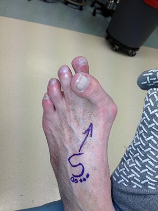
Hallux valgus is one of the most recognizable deformities of the foot. (See Figures 3-7.) Investigators consistently have reported that this disorder is more common in females than males; however, this finding may be inaccurate, since the studies cited were based on relatively small numbers of subjects.22 Women are more likely than men to consult with their primary care physicians. The true prevalence of hallux valgus in the general population remains unknown. Many factors, including foot type, biomechanical instability, shoe gear, and other hereditary factors, affect the development of this deformity. Women generally have more issues with foot gear, thus giving the appearance that symptomatic hallux valgus deformity occurs more in women. 23
Figure 3. Clinical Photograph of a Bunion

Figure 4. X-Ray of a Bunion
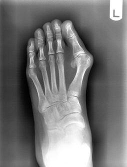
Figure 5. Clinical Photograph of a Postoperative Bunion Correction
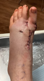
Figure 7. Clinical Photograph of a Bunion
Hallux valgus is a progressive subluxation of the first metatarsal phalangeal joint. The normal anatomic variant is slight lateral deviation in the great toe with a hallux valgus angle of less than 15 degrees. Mild deformities exhibit an angle of 20 degrees, with moderate deformities at 30 degrees and severe deformities more than 40 degrees. As the hallux moves laterally, retrograde force is placed proximally on the first metatarsal head. The medial and dorsal medial aspect of the first metatarsal becomes “prominent,” thus leading to the bunion formation. Over time, uneven degeneration of the first metatarsal phalangeal joint can lead to arthritis. It is important for the clinician to be able to differentiate the etiology of patient’s symptoms.
The presenting symptoms include pain in the medial aspect of the metatarsal phalangeal joint, with redness and swelling at the area of excessive shoe pressure. There may or may not be pain with range of motion of the metatarsal phalangeal joint. The range of motion can be mildly decreased.
Initial treatment depends on the severity of the deformity. In mild cases, wider or larger shoes will give adequate relief. If there is a concomitant bursitis of the joint, nonsteroidal anti-inflammatory medications may be of great benefit in the short term. Various types of padding may be used to cushion the medial joint space. Foot orthoses can support and offload the first metatarsal phalangeal joint to take weight off the area. Some patients may present with increased pain with a mild or moderate deformity, which can be explained by an entrapment of the medial saphenous nerve.
Surgery can be beneficial in hallux valgus deformities that are painful and in the moderate to severe range. There are many surgical procedures described in the literature, but the goal is to reduce the hallux valgus angle and to relocate the first metatarsal phalangeal joint to a more anatomic position. Surgeries include soft tissue balancing of the first metatarsal phalangeal joint combined with osteotomies of the great toe and first metatarsal to realign the toe. Sometimes, an arthrodesis of the first metatarsal tarsal joint is required to address any instability of that joint to stabilize and reduce the first intermetatarsal angle to an anatomic position.
Hallux Limitus and Hallux Rigidus
Hallux limitus is a condition in which there is limited dorsiflexion of the first metatarsal phalangeal joint to less than 20 degrees. Hallux rigidus is the end stage of hallux limitus, when the joint undergoes degenerative changes. A common presenting symptom is pain with limited range of motion. Hallux limitus has many causes. Multiple factors influence the loss of motion in the joint. Some causes include acute trauma, arthritis of the sesamoids, abnormal subtalar joint pronation, long first metatarsal, long proximal phalanx of the great toe, metatarsus primus elevatus, square first metatarsal head, and impingement of the sesamoid apparatus and flexor hallucis longus tendon in the retromalleolar region.
Hallux rigidus is the second most frequent site of arthrosis, after the knee.24 There are few or no preventive measures readily available. Symptomatic treatment includes offloading the first metatarsal phalangeal joint and limiting abnormal biomechanical forces with foot orthotics, shoe modifications (including a show with a stiffer sole or one with a rocker bottom), nonsteroidal anti-inflammatory medications, short-acting corticosteroid intra-articular injections, and physical therapy. Surgeries include cheilectomy or debridement of the first metatarsal phalangeal joint of ankylosed bone, implant arthroplasty, and various osteotomies of the proximal phalanx or first metatarsal to decompress the joint. Salvage procedures include arthrodesis of the first metatarsal phalangeal joint. Early detection and diagnosis are paramount to limiting the progression of this disorder.
Digital Contractures
Typical deformities affecting the lesser toes include hammertoe and claw toe deformities. These deformities are reported in the literature to affect 20% of the population.25 Surgeries for these deformities are among the most commonly performed procedures in foot and ankle practices.
Pathologies regarding contracted pedal digits result from muscle imbalances that occur both from a pronated or flat foot and from a supinated or high arched foot. The mechanism of deformity among hammertoes, claw toes, and mallet toes is different, but the result can appear similar. Subsequently, contracted digits affect the distribution of pressure across the weight-bearing surface of the foot. The heads of the five metatarsal bones are weight-bearing structures, and any toe contracture will place retrograde pressure on the metatarsal heads, which often leads to painful conditions.26 These manifest in different ways, including calluses, capsulitis, and metatarsalgia. The conditions are not mutually exclusive and often present with multiple symptoms. It is important to identify the cause of symptoms to alleviate the pain and prevent long-term progression.
A “true” hammertoe is characterized by dorsiflexion at the metatarsophalangeal joint and plantarflexion of the proximal interphalangeal joint, then dorsiflexion at the distal interphalangeal joint. A claw toe occurs when a contracture is present at both the proximal and distal interphalangeal joints.
Clinically, the metatarsophalangeal joint dorsiflexion results in the head of the proximal phalanx to push against the internal surface of shoes, exerting abnormal pressures on the head of the proximal phalanx. Abnormal contractures at the proximal interphalangeal and distal interphalangeal joints cause abnormal skin irritation, creating painful hyperkeratoses, nail disorders, and ulcerations.
Conventional conservative care includes sharp debridement of the clavus along with accommodative padding, larger shoes, or a change in shoes to ones with a deeper toe box. Caustic callus removers should be discouraged because of the potential for severe skin burns, especially in the neuropathic foot. If the digital deformities are flexible and there are signs of abnormal foot mechanics, foot orthotics may be beneficial.25
Complex hammertoe deformities exhibit biplane instability with dorsiflexion of the toe and either adduction or abduction at the metatarsophalangeal joint. If this occurs in the second toe, it typically is referred to as a classic overlapping toe. It can present with a hallux valgus deformity. This is a much more difficult type of hammertoe to treat surgically.27
In advanced digital deformities, surgery often is indicated with resectional arthroplasty of the affected proximal interphalangeal joint and distal interphalangeal joint. Arthrodesis of the proximal interphalangeal joint is used as a more stabilizing procedure. In the case of painful, semi-rigid hammertoes or toes with distal hyperkeratoses that exhibit potential ulceration, a simple flexor tenotomy may be beneficial for relieving pressure from the distal end of the toe. This is especially useful in neuropathic patients with digital deformities to prevent recurrent ulcerations.5
Metatarsalgia, Capsulitis, and Morton’s Neuroma
Metatarsalgia, capsulitis, and Morton’s neuroma are grouped together because they usually are differential diagnoses of one another. These three conditions are harder to differentiate, and the history and physical exam are critical in proper diagnosis.28
Metatarsalgia is a nonspecific term used to describe pain along the plantar aspect of the lesser metatarsal heads. Increased and uneven pressure under the metatarsal heads is thought to be the causative factor. Lesser metatarsal overload is thought to lead to a variety of pathologies, ranging from isolated metatarsal pain to complete dislocation of the metatarsophalangeal joint. Initial plantar metatarsal head pain may be a precursor to dislocation of the metatarsophalangeal joint.
Capsulitis or metatarsophalangeal joint synovitis of the lesser metatarsal heads is characterized by localized edema and inflammation plantarly; however, there may be periarticular edema noted dorsally. A less common differential diagnosis in this case may represent early osteochondrosis of the metatarsal head.
Morton’s neuroma is a painful condition that affects the intermetatarsal spaces. Typically, it affects the second or third intermetatarsal spaces. Its cause is the subject of debate. Currently, pathophysiology is thought to be from either a degenerative process or damage to the common digital nerve resulting from ischemia. Symptoms in its early stages present similarly to metatarsalgia. A positive Mulder’s sign (clicking with lateral compression of the forefoot) is not always present and is not pathognomonic for this condition, although it is present in a large number of cases. Pain presents on palpation of the affected intermetatarsal space. Usually, there is no pain on palpation of the metatarsal head. Deliberate and thorough palpation of the forefoot is critical in diagnosing this condition. Typically, this condition worsens with wearing shoes, and removing shoes relieves the pain. Symptoms commonly are described as a burning or stabbing radiating pain to the toes. Radiographic evaluation of the foot is necessary to assess any early signs of metatarsophalangeal subluxations or contractures.
The most common ways to treat these conditions are with metatarsal padding and foot orthotics. Intra- or periarticular corticosteroid injections can exacerbate these conditions by causing rupture of the periarticular capsular ligaments. Although corticosteroid injections may relieve neuroma symptoms, they should be used judiciously and infrequently. Oral anti-inflammatory medications have limited efficacy in treating neuromas but can be quite useful for capsulitis or metatarsalgia. Wider shoes play a significant role in treating interdigital neuromas.
Neuropathy
Peripheral neuropathy involves a wide range of pathologies encompassing peripheral nervous system dysfunction.29 It can present with both positive symptoms such as burning, pain, hyperalgesia, and tingling, to negative symptoms, such as numbness. Metabolic disorders, such as diabetes, are the predominant etiology of peripheral neuropathy; however, numerous others exist, such as a history of chemotherapy, increased alcohol consumption, vitamin or electrolyte deficiencies, or musculoskeletal abnormalities, such as spinal stenosis.
A comprehensive clinical exam is paramount in evaluating the most likely cause of each patient’s neuropathy. The most common cause, diabetes, eventually can lead to serious complications, such as loss of limb and loss of life. Early assessment of risk factors and nerve status is critical to reduce morbidity and mortality resulting from neuropathic foot ulcers and possible underlying pathophysiologic poor wound healing potential.
Peripheral neuropathy largely is diagnosed by a thorough examination, including temperature sensation, vibratory sensation, and light touch sensation via a 10-g monofilament.
First- and second-line therapy options mostly entail proper diet and exercise in addition to optimization of blood glucose, particularly in patients with diabetes. Pharmacologic therapies, such as gapapentinoids (pregabalin and gapapentin), antidepressants, or topical anesthesia medications, also are often indicated. More novel approaches include electrical stimulation therapy.30 It has been shown that an appreciable reduction in pain can be achieved with the use of electrical stimulation. The physiologic effects of electrical current applied to cutaneous nerves are widely unknown, but it is thought that over time this stimulation can increase permeability of nerve cells to nutrients and stimulate increased perfusion to axons. This treatment could support a healthier physiologic environment to allow for peripheral nerves to function more efficiently and subsequently reduce symptoms.
Metatarsal Stress Fractures
The most frequent location of stress fractures in the foot is in the metatarsals.31 (See Figure 8.) The second and third metatarsals are affected the most along the distal shaft and neck region of the metatarsal bone. Stress fractures result from a sudden increase in physical activity or from repetitive stress on the affected area. Initial signs and symptoms are pain, localized edema, and, sometimes, an increase in warmth and localized redness along the dorsal distal part of the foot.
Figure 8. Stress Fracture
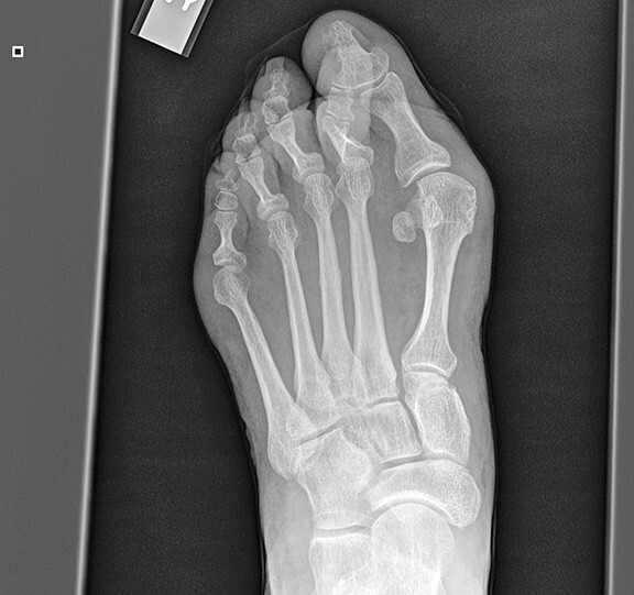
Initial X-rays may be negative, and it may take several weeks to see the callus formation at the fracture site on subsequent X-rays. If initial X-rays are negative (which is common), applying a tuning fork to the appropriate metatarsal will be painful to the patient, indicating movement in the cortical margins of the bone. This is a good indicator that the patient has a stress fracture.32 However, if in doubt, magnetic resonance imaging or technetium Tc 99 bone scan may be performed to help in the diagnosis.
Treatment consists of the PRICE (protection, rest, ice, compression, elevation) protocol through offloading in either a very stiff-soled athletic shoe or surgical shoe. The fracture usually will heal in six to eight weeks. Physical activities should be moderated until symptoms abate.
Conclusion
Many foot problems can be averted with proper shoe gear and patient education. Foot type and biomechanical instability are the root causes of many foot and ankle problems. Even proximal joint pain and lower back pain can be improved if the foot is evaluated for any structural instability. The foot is the foundation for the rest of the body, and proper and timely diagnosis can alleviate a lifetime of problems.
Kevin F. Sunshein, DPM, Fellow, American College of Foot and Ankle Surgeons; CEO, Sunshein Podiatry Associates, Centerville, OH
Carl Braunschweiger, DPM, American Board of Foot & Ankle Surgery; Diplomate, American Board of Podiatric Medicine; Sunshein Podiatry Associates, Centerville, OH
References
- Netter FN. Section 8: Lower Limb Ankle and Foot. In: Atlas of Human Anatomy. Elsevier;2018:515-528.
- Root ML, Weed JH, Orien WP. Normal and Abnormal Function of the Foot: Clinical Biomechanics. Clinical Biomechanics Corp;1977.
- Ficke J, Byerly DW. Anatomy, Bony Pelvis and Lower Limb: Foot. StatPearls [Internet]. Updated Aug. 7, 2023. https://www.ncbi.nlm.nih.gov/books/NBK546698/
- Bonavilla EJ. Histopathology of the heloma durum: Some significant features and their implications. J Am Podiatry Assoc. 1968;58(10):423-427.
- Laborde JM. Neuropathic toe ulcers treated with toe flexor tenotomies. Foot Ankle Int. 2007;28(11):1160-1164.
- Witchey DJ, Witchey NB, Roth-Kauffman MM, Kauffman MK. Plantar warts: Epidemiology, pathophysiology, and clinical management. J Am Osteopath Assoc. 2018;118(2):92-105.
- Wright PK, Vidyadharan R, Jose RM, Rao GS. Plantar verrucous carcinoma continues to be mistaken for verruca vulgaris. Plast Reconstr Surg. 2004;113(3):1101-1103.
- Chen J, Wu Y. Local Treatments for Cutaneous Warts. In: Williams HC, Bigby M, Herxheimer MB, et al, eds. Evidence-Based Dermatology. John Wiley & Sons;2014:320-328.
- Ronna T, Lebwohl M. Cimetidine therapy for plantar warts. J Am Podiatr Med Assoc. 1995;85(11):717-718.
- Hagon W, Hagon J, Noble G, et al. Microwave therapy for the treatment of plantar warts. J Foot Ankle Res. 2023;16(1):37.
- Leyden JL. Tinea pedis pathophysiology and treatment. J Am Acad Dermatol. 1994;31(3 Pt 2):S31-S33.
- Scher RK, Rich P, Pariser D, Elewski B. The epidemiology, etiology, and pathophysiology of onychomycosis. Semin Cutan Med Surg. 2013;32(2 Suppl 1):S2-S4.
- Lipner SR, Scher RK. Onychomycosis: Treatment and prevention of recurrence. J Am Acad Dermatol 2018;80(4):853-867.
- Haneke E. Controversies in the treatment of ingrown nails. Dermatol Res Pract. 2012;2012:783924.
- Espensen EH, Nixon BP, Armstrong DG. Chemical matrixectomy for ingrown toenails: Is there an evidence basis to guide therapy? J Am Podiatr Med Assoc. 2002;92(5):287-295.
- Crawford F, Thomson C. Interventions for treating plantar heel pain. Cochrane Database Syst Rev. 2003;(3):CD000416.
- The Podiatry Institute; Southerland J, Alder D, Boberg J, et al, eds. McGlamry’s Comprehensive Textbook of Foot and Ankle Surgery. Wolters Kluwer Health/Lippincott Williams & Wilkins;2013:494.
- Johal KS, Milner SA. Plantar fasciitis and the calcaneal spur: Fact or fiction? Foot Ankle Surg. 2012;18(1):39-41.
- Schon LC, Glennon TP, Baxter DE. Heel pain syndrome: Electrodiagnostic support for nerve entrapment. Foot Ankle. 1993;14(3):129-135.
- Rufai A, Ralphs JR, Benjamin M. Structure and histopathology of the insertional region of the human Achilles tendon. J Orthop Res. 1995;13(4):585-593.
- Gould JS. Chapter 33: Haglund Deformity, Retrocalcaneal Bursitis and “Pump Bumps.” In: The Handbook of Foot and Ankle Surgery: An Intellectual Approach to Complex Problems. Jaypee Brothers Medical Publishers (P) Ltd.;2013:205-207.
- Chang TJ. Master Techniques in Podiatric Surgery: The Foot and Ankle. Lippincott Williams & Wilkins;2005.
- Nix S, Smith M, Vicenzino B. Prevalence of hallux valgus in the general population: A systematic review and meta-analysis. J Foot Ankle Res. 2010;3:21.
- Coughlin MJ, Shurnas PS. Hallux rigidus: Demographics, etiology, and radiographic assessment. Foot Ankle Int. 2003;24(10):731-743.
- [No authors listed]. Hammer toe syndrome. American College of Foot and Ankle Surgeons. J Foot Ankle Surg. 1999;38(2):166-178.
- Myerson MS, Shereff MJ. The pathological anatomy of claw and hammer toes. J Bone Joint Surg Am. 1989;71(1):45-49.
- Coughlin MJ. Lesser-toe abnormalities. Instr Course Lect. 2003;52:421-444.
- Clinical Practice Guideline Forefoot Disorders Panel; Thomas JL, Blitch EL 4th, Chaney DM, et al. Diagnosis and treatment of forefoot disorders. Section 2. Central metatarsalgia. J Foot Ankle Surg. 2009;48(2):239-250.
- Bodman MA, Dreyer MA, Varacallo MA. Diabetic Peripheral Neuropathy. StatPearls [Internet]. Updated Feb. 25, 2024. https://www.ncbi.nlm.nih.gov/books/NBK442009/
- Dubinsky RM, Miyasaki J. Assessment: Efficacy of transcutaneous electric nerve stimulation in the treatment of pain in neurologic disorders (an evidence-based review): Report of the Therapeutics and Technology Assessment Subcommittee of the American Academy of Neurology. Neurology. 2010;74(2):173-176.
- Clinical Practice Guideline Forefoot Disorders Panel; Thomas JL, Blitch EL 4th, Chaney DM, et al. Diagnosis and treatment of forefoot disorders. Section 5. Trauma. J Foot Ankle Surg. 2009;48(2):264-272.
- Schneiders AG, Sullivan SJ, Hendrick PA, et al. The ability of clinical tests to diagnose stress fractures: A systematic review and meta-analysis. J Orthop Sports Phys Ther. 2012;42(9):760-771.