ECG Review-Chest Pain and Lots of P Waves
August 15, 2001
ECG Review-Chest Pain and Lots of P Waves
By Ken Grauer, MD
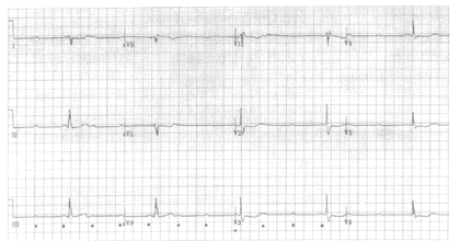 |
| Figure. 12-lead ECG obtained from a 55-year-old woman with chest pain and lots of P waves |
Clinical Scenario: The 12-lead ECG shown in the Figure was obtained from a 55-year-old woman with new-onset chest pain. Many more P waves than QRS complexes are seen on the tracing (see dots under P waves in leads III, aVF, and V3). How would you interpret this ECG?
Interpretation: Although a single lead rhythm strip is lacking, the 12-lead ECG in the Figure can still be interpreted. A narrow-complex marked bradycardia is present that is fairly regular at a rate just over 30 beats/minute. As noted above, many more P waves than QRS complexes are present. The atrial rhythm (marked by the dots) is regular at a rate of between 90-95/minute. Despite the fact that many more P waves than QRS complexes are present, P waves appear to conduct, as evidenced by the presence of a fixed PR interval preceding each QRS complex. This finding rules out the possibility of 3° (complete) AV block, in which there is no relationship between P waves and QRS complexes (P waves "march through" the QRS complex when there is 3° AV block). The rhythm must therefore be some type of high-grade 2° AV block, in this case with 3:1 AV conduction (3 P waves are present for each QRS complex). Although high-grade AV block (in which many if not most P waves fail to conduct) is most often the result of Mobitz II 2° AV block, the lack of consecutively conducted P waves anywhere on this tracing precludes definitive diagnosis. It is important to appreciate that on occasion, the usually less severe Mobitz I (Wenckbeach) form of 2° AV block may also be "high grade," with failure of consecutively occurring P waves to conduct. In such situations, the characteristic picture of progressive PR interval lengthening prior to dropping a beat may not be seen.
Analysis of the remainder of the ECG in the Figure reveals marked right axis deviation (RAD) consistent with a left posterior hemiblock (LPHB) pattern, incomplete right bundle branch block (IRBBB) evidenced by an rsr’ pattern in lead V1, early transition (a relatively tall R wave is present in lead V2), and worrisome ST segment depression in leads I, aVL, and V2-V6. An ECG obtained 1 hour earlier showed ST segment elevation in the inferior leads (which has now resolved). The overall picture in this 55-year-old woman with new-onset chest pain is most consistent with acute evolving infero-posterior infarction. Telemetry tracings over the previous hour revealed clear evidence of Mobitz I (Wenckbach) 2° AV block—which in conjunction with the findings of normal QRS duration and acute inferior infarction strongly suggest that the 2° AV block with 3:1 AV conduction seen here is probably also a manifestation of the Mobitz I (Wenckbach) form of 2° AV block. That said—the bifascicular conduction defect and acute infarction with marked bradycardia are clear indication for emergency pacemaker placement.