ECG Review: An Octogenarian in Heart Failure
September 15, 2007
ECG Review
An Octogenarian in Heart Failure
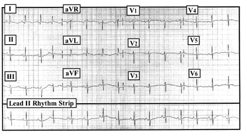 |
| Figure. 12-lead ECG obtained from an 81-year old woman with new onset heart failure, but no chest pain. |
By Ken Grauer, MD, Professor, Department of Community Health and Family Medicine, University of Florida Dr. Grauer is the sole proprietor of KG-EKG Press, and publisher of an ECG pocket brain book.
Clinical Scenario: The ECG in the Figure was obtained from a 81-year old woman who presented with new-onset heart failure. She described shortness of breath, but no chest pain. What may have caused her heart failure?
Interpretation/Answer: The underlying rhythm is sinus at a rate of about 90/minute. There is one PAC (premature atrial contraction), which is the fourth beat. The PR, QRS and QT intervals are normal. The axis is close to 0°. There is no ECG evidence of chamber enlargement. R wave progression is slightly delayed (with transition occurring between leads V4 to V5), and small S waves persist in leads V4 through V6. For the most part, ST-T waves show a flattened appearance that is non-specific. The most interesting part of this tracing (given the clinical context) is seen in the high lateral leads (leads I and aVL). In addition to small q waves, there is subtle (but real!) ST segment elevation. This is clearly the case for all four beats (including the PAC) in lead I, and admittedly less certain (due to beat-to-beat variation) in lead aVL. This patient had positive troponins that confirmed her acute high lateral myocardial infarction.