
Pediatric Tachycardia?
July 15, 2025
By Ken Grauer, MD
The electrocardiogram in the figure is from a previously healthy 15-year-old boy who reports palpitations and dizziness over the past two weeks. How would you interpret this tachycardia?
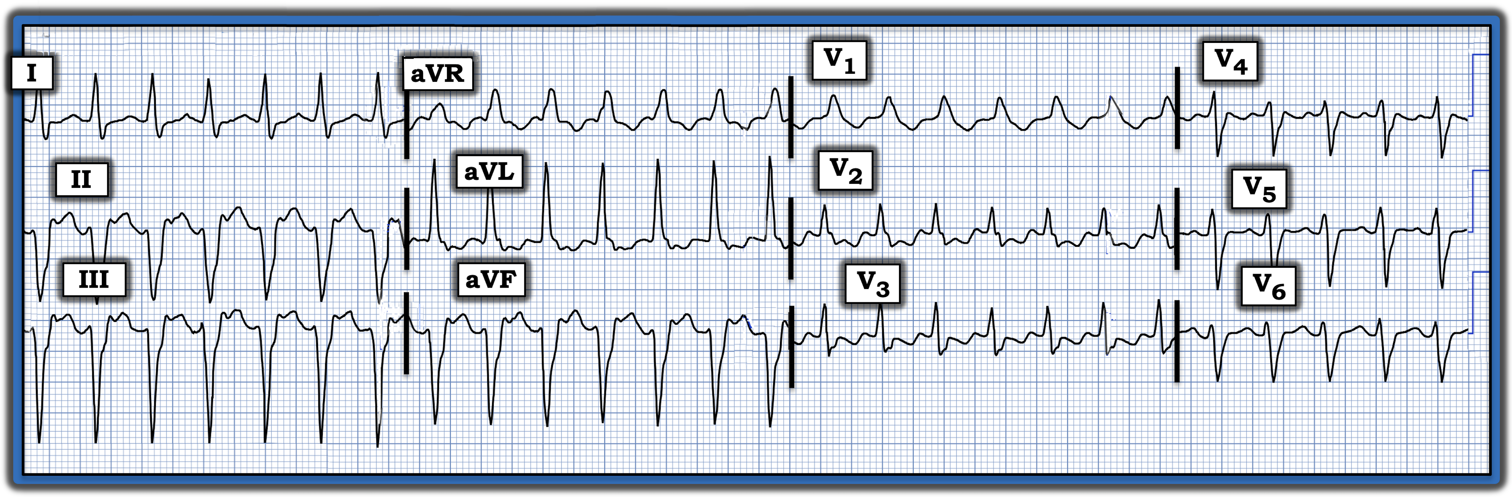
Interpretation: Although there is no long lead rhythm strip in today’s tracing, it is clear that there is a regular wide-complex tachycardia (WCT) at a rate of
~160-170 beats per minute.
- Sinus P waves are absent. That said, there may be indication of 1:1 retrograde P waves in the form of shallow negative deflections following the QRS complex in each of the inferior leads. This is not atrioventricular dissociation because the RP’ interval (distance from the end of the QRS until these negative deflections) is constant. Since both ventricular tachycardia and supraventricular tachycardia rhythms may manifest retrograde conduction — this finding (if real) does not help in the differentiation of today’s WCT rhythm.
QRS morphology in today’s rhythm superficially resembles right bundle branch block (RBBB) with left anterior hemiblock (LAHB).
- That said, the monophasic R wave in lead V1 is atypical for RBBB conduction in a previously healthy adolescent, in whom one would expect to see a triphasic (rSR’) complex. This finding makes aberrant conduction and preexisting RBBB less likely.
- The almost entirely negative QRS complexes in each of the three inferior leads (II, III, aVF) also is atypical for the LAHB pattern expected when there is aberrant conduction, in that the initial small positive (r wave) deflection expected with LAHB in the inferior leads is almost entirely absent.
Impression: The finding of a regular WCT rhythm in a younger patient that superficially resembles a bifascicular block pattern — but is lacking in typical features for RBBB and LAHB — suggests that the rhythm is much more likely to be fascicular ventricular tachycardia (VT).
- Fascicular VT is one of the more common forms of idiopathic VT — in which patients tend to be younger and do not have underlying heart disease. This rhythm often responds to intravenous verapamil, which successfully converted the rhythm to sinus.
For more information about this case, visit https://tinyurl.com/KG-Blog-464.