
And Then What Happens?
May 15, 2025
By Ken Grauer, MD
You are consulted on the electrocardiogram (ECG) in the figure — but without the benefit of any clinical information. This tracing was assessed as showing AFib (atrial fibrillation) with a controlled ventricular response. Do you agree?
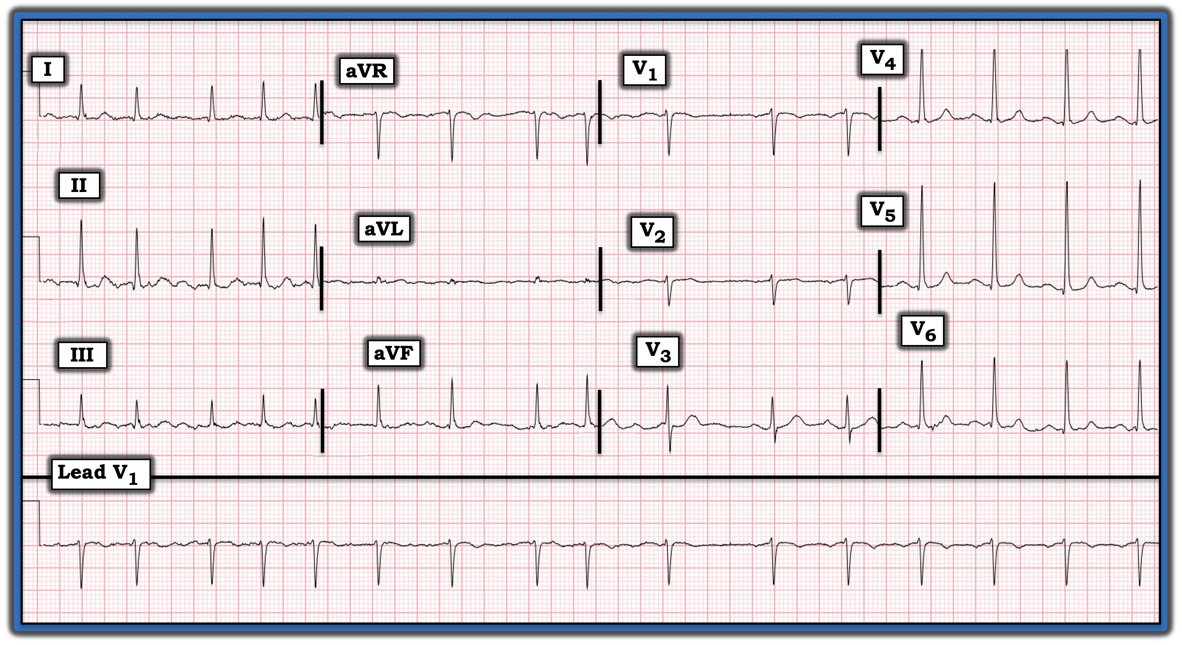
Interpretation: The ECG in the figure displays a simultaneously recorded long lead V1 rhythm strip under the 12-lead tracing. The initial part of this long lead V1 is consistent with AFib in that the QRS complex is narrow, the rhythm is irregularly irregular, and no P waves are seen. The ventricular response is not overly rapid.
A look at simultaneously recorded lead II fails to show any P waves, thereby confirming AFib during the initial portion of this tracing.
That said, the rhythm changes and becomes regular for the last six beats in the long lead rhythm strip.
- Each of these last six beats is preceded by a small amplitude negative P wave deflection with a constant PR interval.
Impression: Today’s ECG is interesting in that it captures spontaneous conversion of AFib to sinus rhythm toward the end of the tracing.
- It is extremely easy to overlook this spontaneous conversion to sinus rhythm because, instead of a long lead II rhythm strip (in which sinus P waves will be upright and usually much easier to detect), lead V1 was selected for the long lead rhythm. And, because sinus P waves often are negative in lead V1 — and the P waves in today’s case are of very small amplitude — initial inspection misinterpreted these very small sinus P waves as “fib” waves.
- Assessment of the 12-lead tracing confirmed narrow QRS complexes, a normal QTc interval, normal frontal plane axis, no chamber enlargement, and no acute ST-T wave changes.
Take-home point: Be sure to look at the entire long lead rhythm strip for assessment of rhythm regularity, atrial activity, and potential relationship between P waves and neighboring QRS complexes. Failure to do so makes it easy to overlook subtle rhythm changes, as occurred in today’s case.
Note: For more information about this case, visit https://tinyurl.com/KG-Blog-466.