Epigenetic Changes in Perilesional Brain Tissue After Radiotherapy
June 1, 2025
By Rajiv S. Magge, MD
Synopsis: Epigenetic and transcriptomic studies of irradiated perilesional brain tissue identified clear changes in deoxyribonucleic acid (DNA) methylation patterns and neuropeptide upregulation that contributed to neuroinflammation, which may underly radiation-related neurotoxicity.
Source: Millner TO, Panday P, Xiao Y, et al. Disruption of DNA methylation underpins the neuroinflammation induced by targeted CNS radiotherapy. Brain. 2025; Apr 29. doi:10.1093/brain/awaf163. [Online ahead of print].
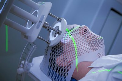
Radiotherapy (RT) remains a central treatment modality for both primary brain tumors and brain metastases. Radiation induces deoxyribonucleic acid (DNA) damage in neoplastic cells, subsequently leading to apoptosis and tumor cell death. Targeted RT via techniques such as stereotactic radiosurgery (SRS) have become a fundamental treatment modality for managing brain metastases and improving survival outcomes. Unfortunately, radiotherapy also can injure adjacent non-neoplastic brain cells, contributing to significant neurotoxicity, such as short-term fatigue, nausea, and headaches, as well as long-term cognitive dysfunction and radiation necrosis, which have limited treatments and often are irreversible.
Many studies have investigated DNA damage and epigenetic changes in irradiated tumor cells, but much is unknown regarding the DNA methylation (DNAme) and transcriptomic changes in surrounding neuronal and glial cells. These changes in the adjacent brain tissue likely contribute to local neuroinflammation and potentially mediate both acute and chronic symptoms of neurotoxicity. Targeting this RT-related epigenetic dysregulation may be a potential treatment avenue to reduce RT-associated neurotoxicity.
Millner et al retrospectively identified 14 neurosurgical samples of perilesional brain tissue within the treatment field of patients who received RT (nine patients were treated for brain metastases). As a control, they also collected 12 samples of perilesional brain tissue surrounding brain metastases from RT-naive patients. Time to resection after RT was quite variable, with a range of seven to 240 months. Histology in the post-RT samples demonstrated prototypical changes, including reactive gliosis, inflammation, vascular changes, and white matter necrosis.
DNAme profiling subsequently was performed on the samples, demonstrating clear methylation changes across the genome of perilesional glial and neuronal cells. Transcriptomic studies also identified microenvironmental niches, with prominent inflammatory cells present in most of the irradiated samples. Several proinflammatory neuropeptides, including tachykinins and opioid precursors, also were highly expressed in irradiated neuronal clusters.
The investigators went a step further and designed a cerebral organoid model of RT, allowing assessment of early neuronal changes after radiation (cerebral organoids were irradiated at day 48 of development with 24 Gy). Epigenetic studies in these cerebral organoid neuronal cells recapitulated similar DNA methylation dysregulation and altered pro-inflammatory neuropeptide expression, as seen in the post-RT patient samples.
Commentary
In this elegant study, Millner et al found that neuronal and glial cells in perilesional brain tissue underwent significant DNA methylation dysregulation associated with elevated pro-inflammatory neuropeptide expression after RT exposure. They cleverly recapitulated similar results in a cerebral organoid model, where early radiation with a standard 24 Gy dose catalyzed clear neuronal epigenetic changes.
There are no effective treatments for RT-associated cognitive dysfunction, and targeting these epigenetic changes in perilesional neuronal and glial cells may represent a path to reduce subsequent neuroinflammation and neurotoxicity. Conclusions remain limited with the low number of samples, which is of course understandable with the challenge of acquiring appropriate samples and the resource-intensiveness of the molecular analyses. Furthermore, the time to resection after radiation was quite variable in the post-RT sample cohort, ranging from seven to 240 months, which represents a significant confounder, especially with the early DNA methylation changes seen after day 48 of radiation in the cerebral organoid neuronal cells.
It also is unclear how different dosing and radiotherapy strategies (such as fractionated intensity-modulated radiation therapy vs. SRS) as well as the diversity of neoplasms being targeted (including secondary brain metastases as well as primary brain tumors, such as glioblastoma) may affect the epigenetic dysregulation seen in cells within the radiation treatment field.
The investigators should be commended on their identification of pathogenic epigenetic and transcriptomic changes that may cause neuroinflammation and mediate RT-induced neurotoxicity, an often-overlooked area of investigation without any clear effective treatment options.
Rajiv S. Magge, MD, is Associate Professor of Clinical Neurology, Weill Cornell Medicine, Weill Cornell Brain Tumor Center.